Skull mesenchymal stem cell (MSC) and skull-brain interface
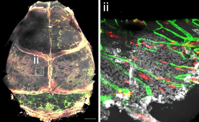
We investigate mesenchymal stem cells (MSCs) residing in the skull and their interactions with specialized niches, including lymphatic endothelial cells (LECs), immune cells, and neurons. Our previous work demonstrated that skull malformation perturbs meningeal lymphatic function and fluid homeostasis, thereby contributing to neurocognitive deficits (Cell Stem Cell, 2023). These findings prompted us to further examine the skull-brain functional interface.
To this end, we employed tissue clearing, high-resolution three-dimensional (3D) imaging, cell type–specific genetic models in mice, and surgical interventions to interrogate skull vasculature. Through these approaches, we characterized the heterogeneity of skull channels, identified lymphatic vessels within the skull periosteum (Video 1), and resolved the longstanding question of whether lymphatic vessels are present within bone tissue (J Exp Med, 2025).
Building on these insights, our current research focuses on elucidating the roles of skull MSCs and their niches, such as LECs, in the pathogenesis and therapeutic targeting of craniofacial disorders, including craniosynostosis and skull injuries.
Temporomandibular joint disorders (TMD) and pain
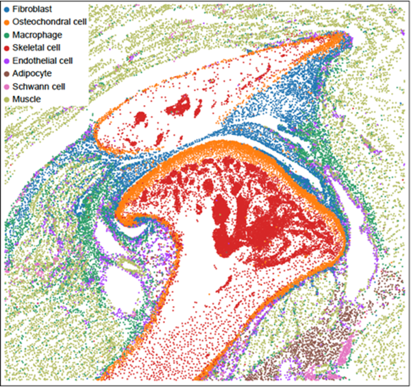
We employed single-cell RNA sequencing (scRNA-seq), spatial transcriptomics (seqFISH), tissue clearing, and three-dimensional (3D) imaging to generate a comprehensive cellular atlas of the temporomandibular joint (TMJ) (JCI Insight, 2025). We functionally mapped nociceptive innervation within the TMJ (Vedio 2) and investigated synovial fibroblast–sensory neuron interactions, the disruption of which contributes to arthritis and pain.
To advance regenerative strategies for temporomandibular disorders (TMD), we integrate lineage tracing, functional assessment, and mechanistic studies to investigate fibrocartilage and tendon/ligament progenitor cells. In parallel, we examine how synovial fibroblasts engage with neurovascular–immune niches to regulate inflammation and pain in TMJ arthritis.
Our research is conducted in collaboration with the multi-institutional Center for TMD Improving Patient-Centered Translational Research (C-TMD IMPACT), facilitating the translation of basic and mechanistic discoveries into improved clinical care and therapeutic interventions for TMD patients.
Lymphatic vasculature and fluid regulation
We established genetic models of small vessel disease by generating SNORD118 mutant mice and human induced pluripotent stem cell (iPSC) lines (Sci Adv; iScience, 2024). Using our craniosynostosis mouse model, we demonstrated that implantation of skull mesenchymal stem cells (MSCs) restores meningeal lymphatic function and fluid homeostasis, thereby improving neurocognitive performance (Cell Stem Cell, 2023).
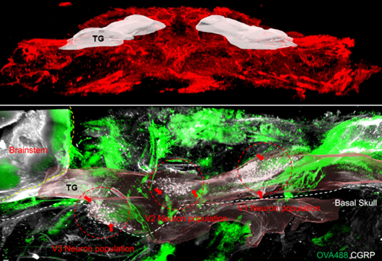
Through whole-brain tissue clearing and high-resolution imaging, we mapped meningeal lymphatic vessels and identified that basal meningeal lymphatics are anatomically proximal to the trigeminal ganglion (TG) (Vedio 3). Cerebrospinal fluid (CSF) flows along trigeminal neurons, enabling uptake of CSF solutes. We discovered distinct meningeal lymphatic–CSF solute exchange loops that modulate trigeminal neuron activation and pain resolution.
Our ongoing research investigates the bidirectional crosstalk between neurons and lymphatic vessels, as well as the interplay between the meningeal lymphatic and glymphatic systems in fluid regulation. These studies encompass a range of pathological contexts, including craniosynostosis, migraine, small vessel disease, and dementia.
Human iPSC cells & organoids for neuro-craniofacial disorders
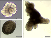
We have established human induced pluripotent stem cell (iPSC)–derived brain and salivary gland organoids, as well as multiple mesenchymal cell types, including suture mesenchymal stem cells (MSCs), fibrocartilage cells, and synovial fibroblasts (Stem Cell Reports, 2023; Sci Adv, 2024). Using CRISPR-based genome editing, we introduce disease-associated mutations into iPSCs or directly derive patient-specific iPSCs from clinical specimens. These platforms enable downstream generation of organoids for FDA-approved drug screening and parallel testing in preclinical animal models.
Our patient-derived and genome-engineered iPSC systems are further integrated with biomaterial and bioengineering strategies to model diseases, investigate pathogenesis mechanisms, develop therapeutic interventions, and identify candidate compounds for conditions such as craniosynostosis, temporomandibular disorders (TMD), and salivary gland–associated xerostomia.